
Molecular sensors
Volume electron microscopy is a rapidly advancing technology that offers nanometer-scale resolution over imaging volumes with edge lengths that are orders of magnitude longer. However, despite the rich ultrastructural information contained in densely labeled EM samples, obtaining multiplexed information about cellular states at EM resolution has been challenging.
EMcapsulins - We have developed a suite of gene reporters for correlated light and electron microscopy (CLEM) that show up as concentric barcodes in EM cross-sections.
Specifically, we have synthesized a series of 6 spherically symmetric barcodes by fusing self-assembling nanocompartments of different sizes derived from the encapsulin family of proteins with different numbers of metal-binding domains (EMcapsulins). These barcodes are readable in TEM and volume SEM.
To further extend multiplexing capabilities, multiple EMcapsulins can also be organized in modular configurations with rigid protein connectors of different lengths.
In addition, EM gene reporters can be made fluorescent and targeted to specific proteins of interest. Using this strategy, we have been able to distinguish genetically defined cells in cell cultures and brains of Drosophila or mice using standard TEM, SEM, and FIB-SEM.
The central constructs listed in Supplementary Table 2 will be distributed via Addgene.
Please get in touch with us if you have any questions about EMcapsulins.
▶︎ Sigmund et al. Nature Biotechnology 2023
▶︎ Westmeyer, Sigmund Nature Biotechnology Research Briefing
in the media:


EXSISERS | exon-specific isoform expression reporter system
Many genes undergo a process called alternative splicing, which results - for coding genes - in the production of different protein isoforms, which serve different functions within cells.
However, until recently, there was no method to monitor the expression of specific protein isoforms in living cells over time and with high throughput.
We thus developed a genetically expressed system called EXSISERS based on a suite of reporter proteins that self-excise themselves from the nascent amino acid chain of a specific isoform being translated from alternatively spliced mature mRNA.
We demonstrated EXSISERS for monitoring the isoform-specific expression of the microtubule-associated protein tau (MAPT), which is involved in neurodegenerative diseases.
in the media:
in the media:
INSPECT | intron-encoded scarless programmable extranuclear cistronic transcript
Although gene reporters are indispensable for biomedical research, they have technically been limited to co-expressing the reporter together with genes that code for proteins.
However, coding genes make up just a small percentage (< 2%) of all human genes.
We tackled this challenge by developing a method to encode reporter or effector proteins from intronic information, i.e., DNA stretches normally spliced out and then degraded. Our method, called INSPECT, instead stabilizes the intronic element, exports it out of the nucleus via specialized export elements, and allows for cap-independent translation.
▶︎ Truong et al. Nat. Cell Biol., 2022.
INSPECT thus enables minimally invasive reporter protein monitoring of the regulation of both coding and non-coding genes and does so without modifying the mature RNA or the resulting protein.

Photoacoustic (optoacoustic) tomography is a non-invasive imaging technology that provides volumetric information on the distribution of photoabsorbers with ultrasound resolution. This is possible through the photoacoustic effect which describes the radiationless conversion of electromagnetic energy into mechanical energy that occurs when chromophores thermoelastically expand upon photoabsorption. With the use of tunable lasers, imaging volumes can be obtained at different wavelengths which allows to spectrally deconvolve photoabsorbing molecules.

Calcium Sensor for Photoacoustics (CaSPA) - We have synthesized a calcium sensor for photoacoustics (CaSPA) that changes its absorbance and thus photoacoustic spectrum upon selective binding to Ca2+ (metallochromism). The compound has a low quantum yield and a high resistance to photobleaching which are optimal properties for photoacoustics. In its esterified form, CaSPA is cell-permeable such that we could use it to image intracellular calcium changes in cells, organoids and in vivo in zebrafish larvae with photoacoustic tomography.
▶︎ Roberts et al. J. Am. Chem. Soc, 2018
in the media:
Based on its semi-cyanine scaffold, red-shifted variants of CaSPA could be synthesized that approach the near-infrared absorbance of, e.g., the heptamethine cyanine dyes that we also work with.
Temporally unmixed MSOT (tuMSOT) - To increase the multiplexing capabilities of gene reporters for multispectral optoacoustic tomography (MSOT), we have introduced the concept of discriminating optoacoustic contrast agents not by color (spectral deconvolution) but instead based on the differences in the characteristic kinetics of contrast agent properties (unmixing in the time domain). We demonstrated this idea on the example of reversibly photoswitchable proteins (RSFPs) whose signal trajectories can be photo-controlled. In this way, the particular temporal pattern can be unmixed from other RSFPs or static background signals even if they have much higher signal amplitudes. We have coined this concept “temporally unmixed MSOT” (tuMSOT).
▶︎ Stiel, Deán-Ben et al. Optics Letters, 2015


Biosynthetic pigments for optoacoustics - As a complementary strategy to using chromoproteins as the basis for genetically controlled optoacoustic contrast, we have been working on the genetically controlled biosynthesis of pigments, which are small-molecular chromophores that are generated by multi-enzymatic processes. We have introduced the biosynthetic pigment violacein as a new photobleaching resistant red-shifted contrast agent for photoacoustics that could, for instance, be used for bacterial tumor tracking.
Designer Melanosome - Melanin is a biosynthetic pigment that is attractive because it is synthesized from tyrosine by a single enzyme and its absorbance spectrum extends into the near-infrared range. However, melanin-containing cells usually store melanin in dedicated organelles called melanosomes, whereas overexpressing tyrosinase in standard cells is toxic which strongly limits its use as a gene reporter for photoacoustic imaging. We have therefore worked on genetically controlled proteinaceous nanocompartments - called encapsulins - that self-assemble in mammalian cells and can selectively encapsulate cargo proteins. We could show that by targeting a version of tyrosinase to the encapsulins, melanin production can be sequestered in the ‘designer melanosomes’, thereby maintaining optoacoustic contrast but strongly reducing toxicity.

Magnetic Resonance Imaging (MRI) is a non-invasive imaging technique that can provide full organism coverage at up to near-cellular resolution. Because of the intrinsic low sensitivity of nuclear magnetic resonance (NMR), it is, however, a challenge to obtain molecular contrast with enough sensitivity. Hyperpolarization techniques are thus a valuable way to increase the signal from molecular MRI contrast agents.
Multi-metal MRI Sensor - Dissolution Dynamic Nuclear Polarization (DNP) is an attractive method to increase the signal in NMR/MRI by hyper-polarizing nuclear spins. This occurs via transfer of spin polarization from electrons to nuclei through microwave irradiation of the compound together with a radical at low temperatures.
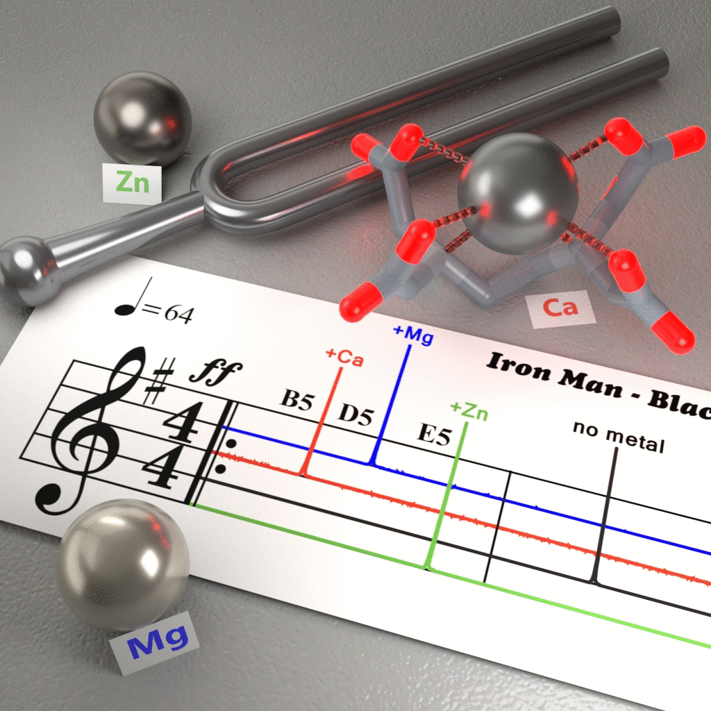
We have developed a sensor based on the well-characterized chelators EDTA and EGTA by introducing a carbon-13 moiety close to the metal-coordination site. The resulting compounds exhibit (1) metal-specific chemical shifts that differentiate several biologically important, or toxic metals, and (2) be hyperpolarized to enable detection of micromolar concentrations of calcium, as well as one-shot metal-specific MR imaging.
Molecular actuators
To understand distributed biological networks and invent effective molecular therapies, we not only require molecular sensors for non-invasive imaging of molecular processes across entire living organ(ism)s. We also need molecular actuators that allow us to manipulate those processes remotely. Ideally, a closed-loop combination of these molecular devices would then enable certain spatiotemporal control of cellular processes to dissect cellular network function and prepare future imaging-controlled molecular therapies.

Magnetic nanocompartments - We have genetically encoded nanocompartments that can biomineralize iron-oxide in their lumen such that mammalian cells expressing them can be visualized by MRI and magnetically sorted by magnetic gradient fields.
These non-toxic and bioorthogonal nanocompartments of 32 nm diameter self-assemble from 180 proteinaceous subunits called encapsulins and harbor ~0.5 nm large pores that allow for the exchange of small molecules. Cargo proteins such as enzymes can be specifically targeted to the inside of the nanocapsules by appending a short encapsulation signal. Excess cargo can be degraded to generate a clean population of encapsulated cargo proteins.
In addition to generating MRI signal, cells expressing the iron-accumulating nanocapsules can be magnetically sorted using commercial sorting columns.
Furthermore, the iron biomineralization inside the capsules also substantially enhances the contrast of encapsulins in electron microscopy (EM) images rendering them excellent genetically encoded EM markers.
In general, we think that genetically encoded orthogonal compartments constitute a powerful generalizable tool for mammalian cell engineering.
in the media:
Genetically controlled delivery of magnetoferritin - In this project, we developed a genetically controlled uptake mechanism for magnetoferritin as a biogenic superparamagnetic nanoparticle. For this purpose, we genetically expressed several ferritin transporters and found that Tim-2 most effectively transports magnetoferritin (made from H-chain ferritin) into the lysosomes of cells in culture.
Lysosomal delivery of magnetoferritin enabled not only robust detection of genetically defined cells by MRI based on the much higher relaxivity of magnetoferritin as compared to regular ferritin. Magnetoferritin also allowed for imaging by optoacoustic and third-harmonic-generation (THG) microscopy because of the strong photoabsorbing properties of ferritin and its lysosomal accumulation.
Interestingly we also found by vibrating sample magnetometry (VSM) analysis, that the blocking temperature of magnetoferritin in the lysosomes of the cells increased dramatically. This change in the magnetic property enabled the application of magnetic hyperthermia which we used to selectively ablate genetically defined cells.
We think that this study showcases an interesting example of a productive interaction between nanoparticles and the cellular environment and identifies magnetoferritin as an interesting candidate for further in vivo work.

Neurobehavioral Imaging
We work with rodent and zebrafish models to study distributed brain functions combining dynamic MRI, optoacoustic and fluorescence imaging with our newly developed molecular devices.
We are in particular interested in studying behaviorally relevant neuronal processes during simple orientation behavior. To enable this work, we develop imaging techniques that allow for capturing molecular information in freely behaving animals.
Neurobehavioral tracking microscope: NeuBtracker - It is a long-standing goal of neuroscience to observe neuronal activity within an entire vertebrate animal while it is freely performing behaviors. Towards this goal, we have built the first tracking microscope for simultaneous imaging of brain activity together with the unperturbed behavior of reporter zebrafish. Using this imaging system, one can, for instance, conduct pharmacological screens for neuroactive drugs by combining behavioral readout and whole-brain molecular neuroimaging, a capability that is currently not possible in any other vertebrate preclinical model system. We are also interested in utilizing NeuBtracker to study orientation behavior during different motivational states.
▶︎ Symvoulidis et al. Nat. Methods 2018

in the media:
Light-independent magnetoreception in zebrafish and medaka - We have conducted behavioral imaging studies that identified zebrafish and medaka fish as genetically tractable and optically accessible model organism to study the biophysical basis of light-independent magnetoreception, the ability to sense Earth’s magnetic field. This is of interest because light-independent magnetoreceptive behavior predicts the presence of magnetic magnetoreceptor cells that convert a magnetic gradient into a mechanical force which in turn leads to a neuronal signal (so-called magnetite hypothesis).
Magnetoreceptive behavior (in the absence of short-wavelength light) can now be confirmed in cohorts of zebrafish and medaka fish and then used to search for the putative magnetic magnetoreceptor cells to elucidate a magneto-mechanical mechanism for magnetoreception. If the putative magnetic magnetoreceptor cells could indeed be found, they could potentially be reverse-engineered to identify essential genetic components of their machinery that may then be used to complement bottom-up synthetic biology approaches that can render cells magnetic under genetic control.
The optically accessible genetic model systems may also help to map the neuronal representation of magnetoreceptive behavior that may ideally be studied during navigation behavior as may be possible using devices such as NeuBtracker.

in the media:
Funding
We are very grateful for the generous support from the following funding sources:


Teaching


Please have a look at the exciting projects that the students of the Munich iGEM teams '16-'18 realized:

Team 2016


Team 2019
